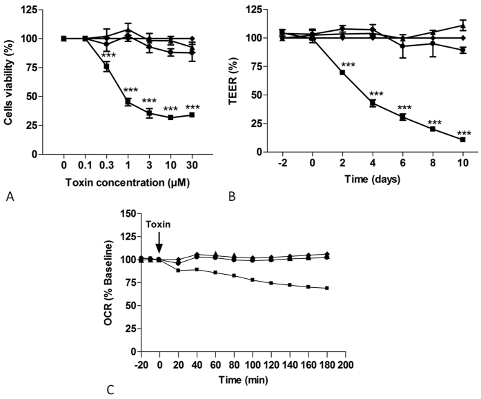
Figure 1. Effects of deepoxy-DON or 3-epi-DON on human intestinal epithelial cells.
Activation of cytotoxicity (Panel A), TEER (Panel B) and OCR (Panel C). (Panel A): Proliferative Caco-2 cells were incubated with increasing concentrations of diluent (♦), DON (◾), deepoxy-DON (▴) or 3-epi-DON (●) for 48 hours. Cell viability evaluated by measurement of ATP, is expressed as % of control cells. (Panel B): Caco-2 cells, differentiated on inserts, were treated with 10 μM of diluent (♦), DON (◾), deepoxy-DON (▴) or 3-epi-DON (●) and TEER was measured. (Panel C): After establishment of baseline oxygen consumption rate in proliferated Caco-2 cells seeded to 1.5 × 10⁴ cells/well, diluent (♦), DON (◾), deepoxy-DON (▴) or 3-epi-DON (●), was injected at final concentration of 10 μM as indicated by the arrow. The rate of oxygen consumption was then measured for the indicated time. For visual clarity, statistical indicators were omitted from the graph. The OCR values are shown as the percent of baseline for each group. Results are expressed as mean ± SEM of 3–4 independent experiments, ***p < 0.001.

