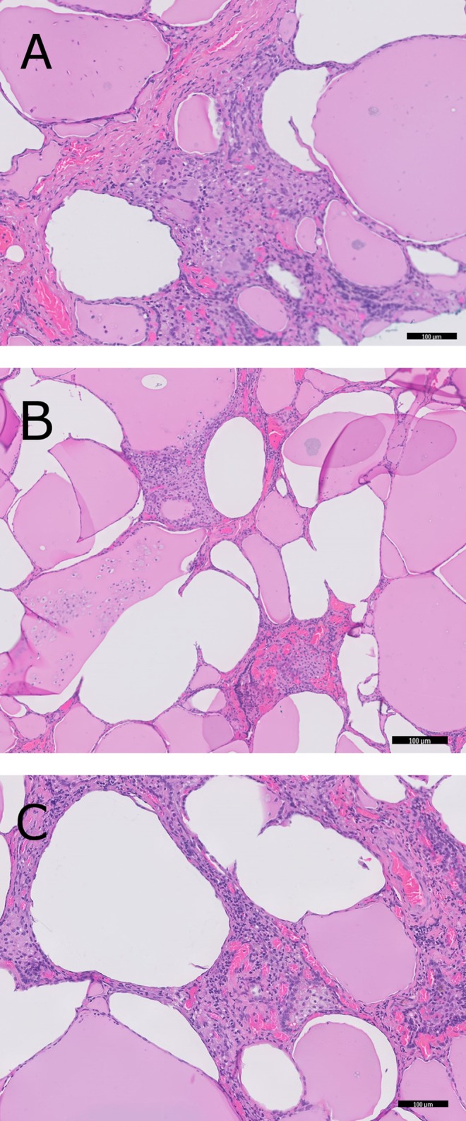
Figure 2.

Microscopic appearance of the thyroid gland. Images are taken at 100× magnification. (A) Follicles with packed stromal tissue containing multinucleated giant cells. (B) Follicles filled with desquamated epithelial cells. (C) Clusters of foamy histiocytes.

 This work is licensed under a