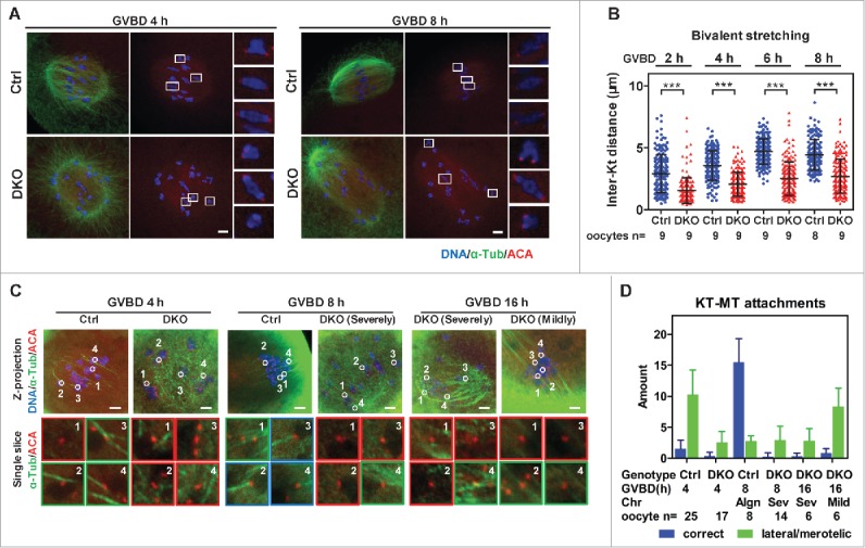
Figure 3.

Lack of bivalent stretching and stable KT-MT attachments in DKO oocytes. (A) Oocytes were immunostained for DNA, α-tubulin and KTs (Anti-centromere antibodies, ACA) to determine bivalent biorientation and stretching. Representative z-projected confocal images of Ctrl and DKO oocytes at GVBD 4 h and GVBD 8 h are shown. Magnified views for 3 representative bivalents in each oocyte are shown in the right side of each z-projected image. Bar = 5 μm. (B) Inter-KT distances of bivalents in DKO and Ctrl oocytes at indicated times after GVBD were plotted (mean ± SD are shown). The numbers of oocytes used for analysis are indicated. Representative result from 3 independent experiments. (C) Oocytes at indicated time points were ice-treated and immunostained for DNA, α-tubulin and KTs (ACA) to determine KT-MT attachments. Representative z-projected images of the whole spindles are shown in the upper row. Magnified views of the KT-MT attachments, indicated by circles and numbers in the upper row, from a single slice, are shown in the lower rows. The types of misalignment of DKO oocytes are indicated. Red panes indicate no attachment; Green panes indicate lateral/merotelic attachments; blue panes indicate correct attachments. Bar = 5 μm. (D) KT-MT attachments in oocytes at times after GVBD were analyzed. Correct attachments (blue column) and lateral/merotelic attachments (green column) were scored (mean ± SD). Chromosome alignments (Algn, aligned; Sev, severely misaligned; Mild, mildly misaligned), numbers of oocytes used for analysis are indicated. Representative result from 3 independent experiments. Statistics: ***, P < 0.001.
