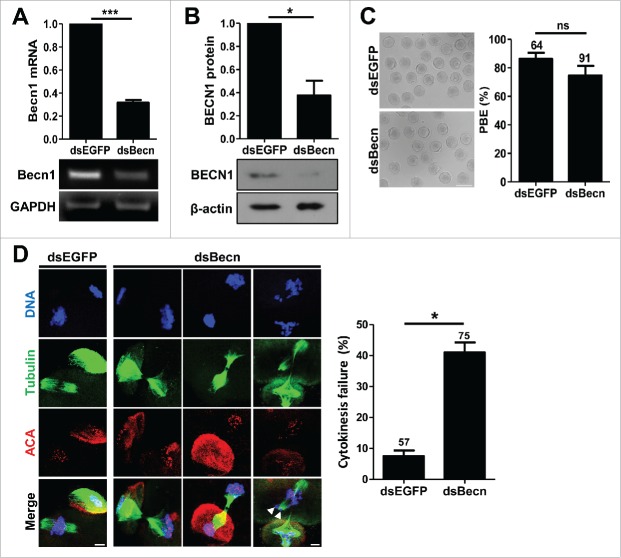
Figure 2.
Knockdown of BECN1 impairs cytokinesis (A, B) GV oocytes injected with indicated dsRNAs were cultured for 24 hours in the presence of IBMX. Knockdown of BECN1 was confirmed by either RT-PCR (A) or immunoblot analysis (B). GAPDH and β-actin were used as a loading control for RT-PCR and immunoblot, respectively. Quantification of the BECN1 level is shown above the representative images. The data are expressed as the mean ± SEM from 3 independent experiments. *p < 0.05; ***p < 0.0001. (C) BECN1 knockdown oocytes were cultured in IBMX-free medium for 13 hours and polar body extrusion (PBE) was scored to assess meiotic maturation. Representative images from at least 3 independent experiments are shown. Bar, 100 μm. The data are expressed as the mean ± SEM. The number of oocytes analyzed was shown above the bar. ns, not significant. (D) BECN1 knockdown oocytes were fixed and stained with anti-tubulin and anti-centromere antibodies (ACA) with DAPI for DNA staining. Representative images from 3 independent experiments are shown. Bar, 10 μm. Arrows indicate the misaligned and lagging chromosomes. The chromosome misalignment and cytokinesis failure were quantified and shown in right panel of images. The data are expressed as the mean ± SEM from 3 independent experiments. The number of oocytes analyzed was shown above the bar. *p < 0.05.

