Figure 1.

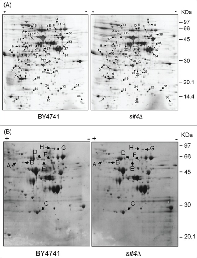
Analysis of changes in the proteome of sit4Δ cells. Yeast extracts were prepared from S. cerevisiae BY4741 and sit4Δ cells grown in YPD medium to exponential phase. Proteins were separated by 2-dimensional gel electrophoresis and visualized by silver staining (A) or blotted into a nitrocellulose membrane. Immunodetection of proteins phosphorylated in serine residues was performed using an anti-phosphoserine antibody (B), as described in Materials and Methods. The experiment was reproduced 3 times, using independent samples. A representative gel/blot is shown. Arrows indicate proteins differentially expressed in sit4Δ cells compared to BY4741 cells.
