
Figure 1.
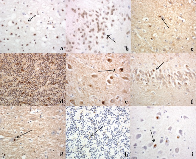
Sparse TDP‐43 pathological changes in case 2, seen as occasional neurites in the cerebral cortex (a), and rare neuronal cytoplasmic inclusions in neurones of the dentate gyrus of the hippocampus (b). Poly‐GA immunostaining shows moderate to many neuronal cytoplasmic inclusions in small neurones of the temporal cortex (c), cerebellar granule cells (d), neurones of CA4 region (e) and granule cells of dentate gyrus (f) of the hippocampus and neurones of the ventrolateral nucleus of the thalamus (g). Immunostaining for p62 protein shows relatively fewer neuronal cytoplasmic inclusions in cerebellar granule cells (h) and CA4 pyramidal cells of the hippocampus (i) compared with poly‐GA immunostaining (compare with (d) and (e) respectively). Immunoperoxidase‐haematoxylin. ×40 microscope objective magnification.