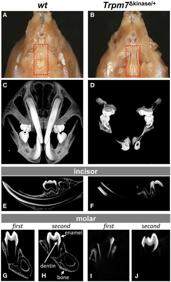
Figure 2.

A hypomineralized phenotype is present in enamel, dentin and cranial bones of Trpm7Δkinase∕+ mice. (A) Transluscent enamel is found in P14 wt mice. (B) The transparent mandibular incisor enamel of P14 Trpm7Δkinase∕+ mice allows the red colored pulp tissue could be seen through. (C) 3-D microCT images of craniofacial structure show well mineralized incisors, entire molars, and craniofacial bones in wt mice. (D) At the same intensity threshold, the mineralized tissues are only detected in the crowns of molars, a small segment of incisors near the incisal end in Trpm7Δkinase∕+. (E) A representive 2-D microCT image of sagittal section from P14 wt mouse hemimandible show the well contrasted enamel, dentin and alveolar bone. (F) The 2-D microCT image of sagittal section from P14 Trpm7Δkinase∕+ hemimandibles show contrasting image intensity consistent with mineralization only seen in the molar crown and incisal end of the incisor. Mineralized enamel, dentin and alveolar bone are detected in P14 wt first molar (G), and second molar (H). Clear contrasts in these tissues is only seen in part of first molar enamel, dentin and alveolar bone (I) and coronal enamel and dentin in the second molar (J) of P14 Trpm7Δkinase∕+ hemimandible.
