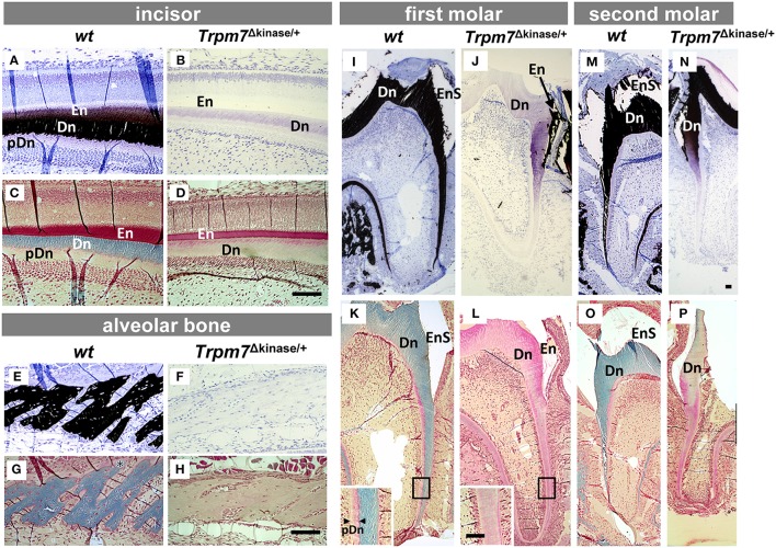
Figure 3.
Hypomineralization defects of enamel, dentin and alveolar bone in Trpm7Δkinase∕+ mice are characterized by histological analyses. Matrix mineralization was assessed by Von Kossa staining (A,B,E,F,I,J,M,N) and Goldner's Trichrome staining (C,D,G,H,K,L,O,P). In the wt incisor (A,C), secretory stage enamel matrix (En) is lighty positive (black) for Von Kossa staining (A), and is stained in red by Trichrome staining (C). Dentin matrix (Dn) is stained in black with Von Kossa (A), and is stained in blue with Trichrome staining (C). Non-mineralized/Von Kossa negative pre-dentin (pDn) is stained in light pink by Trichrome staining (C). In Trpm7Δkinase∕+ mice, both incisal enamel and dentin are negative for Von Kossa staining (B), and Trichrome stains the dentin matrix in light pink, similar to the color of non-mineralized pre-dentin (D). The alveolar bone matrix of wt mice is stained in black by Von Kossa (E), and light blue by Trichrome staining (G). The alveolar bone matrix of Trpm7Δkinase∕+ mice is negative for Von Kossa staining (F), and Trichrome staining on alveolar bone showed bone in Trpm7Δkinase∕+ mice is light pink (H), similar to osteoid. Unlike Von Kossa positive stained wt molar dentin (I,M), the entire dentin of the Trpm7Δkinase∕+ first molar is negative for Von Kossa staining (J) and shows pink/pre-dentin status by Trichrome staining (L). Interestingly, in the second molar of Trpm7Δkinase∕+ mice, both Von Kossa (N) and Trichrome (P) staining shows the coronal dentin as partially mineralized, but the root dentin is not mineralized. Enamel matrix of molars likely chipped off during the sectioning (seen as an enamel space/EnS), and the remaining matrix is positive for Von Kossa staining (N). Scale bars: 100 μm

