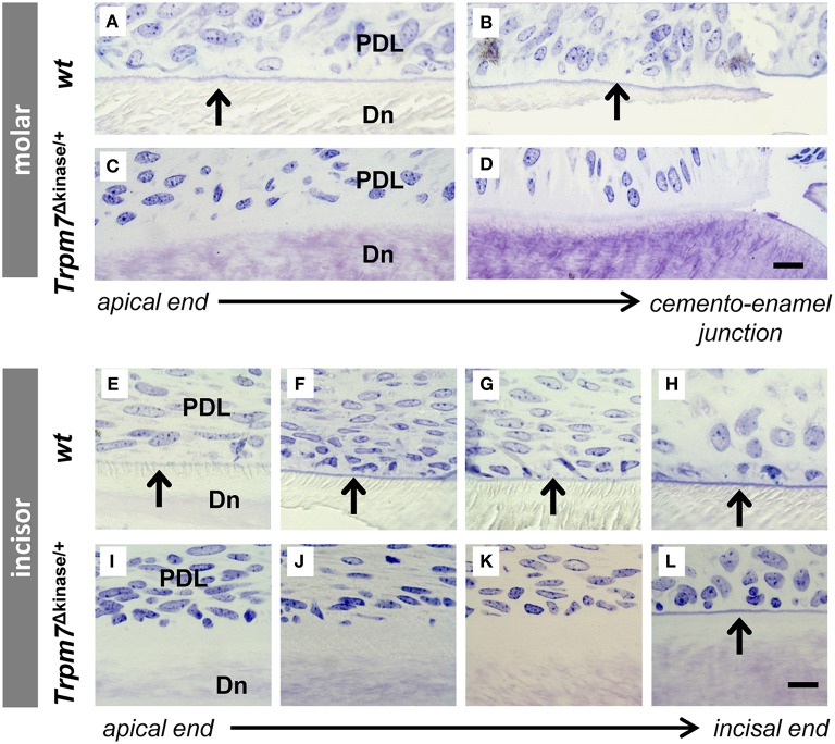
Figure 5.
Acellular cementum is found on the root analog of wt mouse incisor as well as on the molar root. Acellular cementum, ususally seen as a blue line following toluidine blue staining, is visualized between periodontal ligment (PDL) and dentin (Dn) on wt molar (A,B). A similar staining pattern corresponding to acellular cementum can not be detected in Trpm7Δkinase∕+ mice (C,D). Acellular cementum, seen as a blue line by Toluidine Blue staining, is visualized along the incisors from the apical to incisal end in P14 wt mice (E–H, indicated by black arrows). However, this line (indicated by black arrow) only appears on the surface of the incisor root analog dentin near the incisal end in Trpm7Δkinase∕+ mice (I–L). Scale bars: 25 μm

