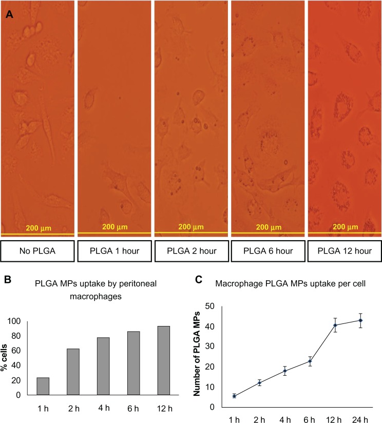
Figure 6.
Uptake of PLGA MPs in vivo. A/J mice were injected with PLGA MPs and peritoneal exudate cells were examined at indicated time intervals (1, 2, 6 and 12 hours post injection) by light microscopy. Shown are representative images for each time point (A), percentage of cells loaded with PLGA MPs (B) and average number of PLGA MPs per cell (C).
Abbreviation: PLGA MPs, Poly lactic-co-glycolic acid microparticles.

