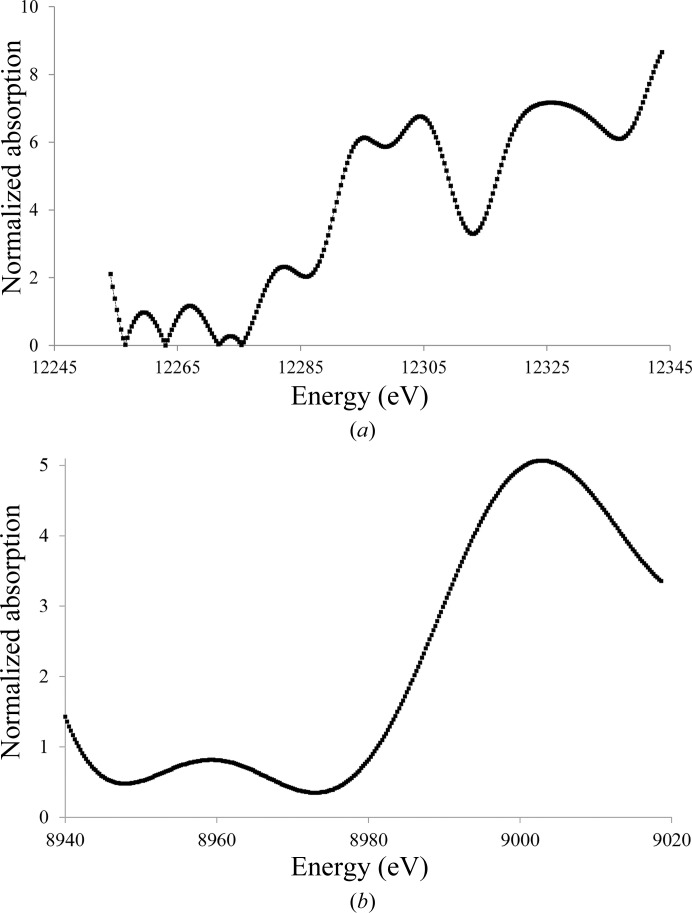
Figure 6.
X-ray absorption spectra of Tth-MCO crystals in the holo form and with Hg bound before X-ray data collection. (a) Hg-Tth-MCO-2h single crystal showing the L III absorption edges of protein-bound Hg. (b) Tth-MCO single crystal showing the Cu K absorption edge of the four native copper(II) atoms of the enzyme.

