
Figure 3. Histological evidence of vascular damage and decreased tumor cell density in 4T1 tumor tissue following combined CYT-6091 and radiation therapy.
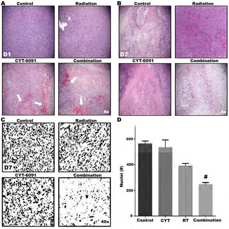
A) Representative H&E images from treatment groups at time of tissue harvest. At day 1, tumor tissue from CYT-6091 and combined therapy groups displays marked vascular hemorrhaging. B) By day 7, tumor cell density in the combined treatment group is markedly reduced. C) Representative digitalized and binarized H&E images from treatment groups at day 7. D) Symbols above bars indicate significant differences resulting from post-hoc statistical tests. Data represent mean number of nuclei ± SEM (#, p<0.05).