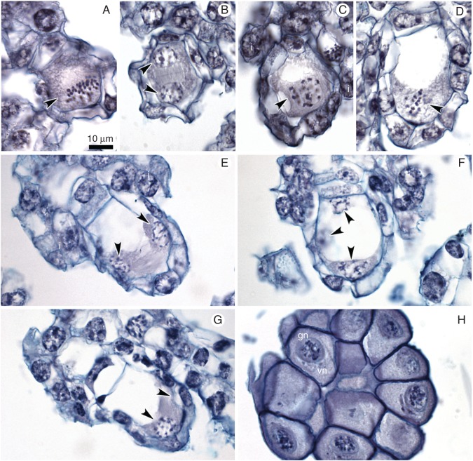
Fig. 4.
Embryological study of the ovules within unpollinated flower buds (control). (A,B) Different stages of meiosis: (A) metaphase I; a group of chromosomes is marked by an arrowhead; integuments (i) are just visible. (B) Late telophase I – two sister nuclei are marked by arrowheads. (C–G) Development of female gametophyte (FG). (C) One-nucleate FG (arrowhead); integuments are visible. (D) Two-nucleate FG; mitotic metaphase at micropylar end of FG (arrowhead). (E–G) Nuclei of FG are indicated by arrowheads; note the lack of cellularization and cell specification. (H) Two-nucleate pollen grains in pollinium; vn – vegetative nucleus, gn – generative nucleus. Scale bar = 10 μm.

