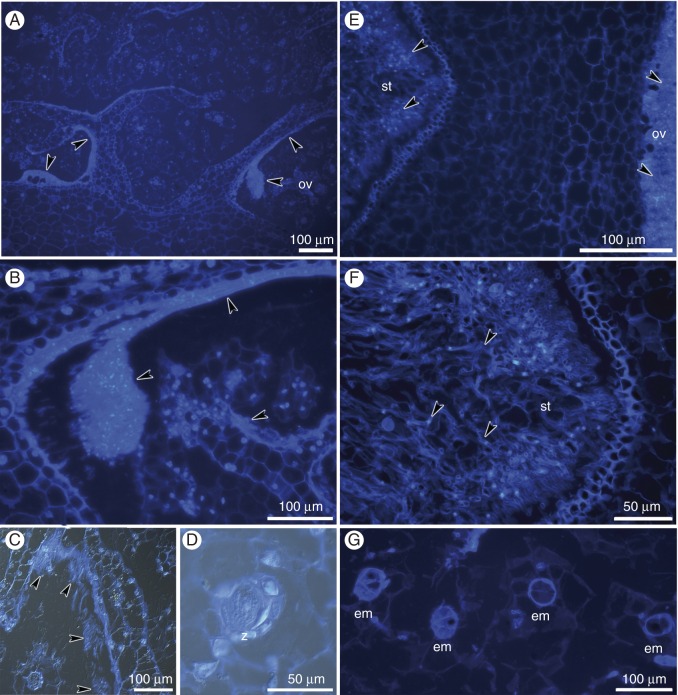
Fig. 7.
Histochemical analysis of hand-pollinated pistils on the 12th (A–D) and 16th (E–G) days after hand-pollination; longitudinal sections of the ovary. (A–G) Chromatin of the nuclei was stained with DAPI, and cell walls were stained with calcofluor white. (C,D) Method combining fluorescence and differential interference contrast. (A–C) Masses of pollen tubes within the ovary (arrowheads). (D) The zygote (z). (E,F) Masses of pollen tubes (arrowheads) within the stylar tissue and the ovary. (G) Two- and four-cell embryos (em).

