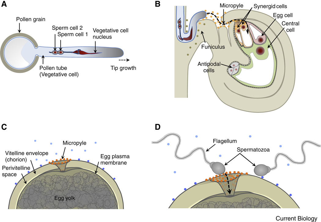
Figure 1. Comparison of gamete/gametophyte morphology between the model plant Arabidopsis thaliana and the model animal Zebrafish.
(A) Diagram of the haploid male gametophyte (pollen) of Arabidopsis comprising the vegetative cell (producing the growing pollen tube) and two non-motile sperm cells enclosed within the a membrane of the vegetative tube cell. The sperm cells are connected to each other and to the nucleus of the vegetative pollen tube cell forming the "male germ unit". Nuclei in red, vegetative cell membrane in blue, sperm cell membranes in black. (B) A pollen tube approaching the Arabidopsis ovule. The tube grows through the micropyle of the ovule along the funiculus towards the haploid female gametophyte that comprises the egg cell, central cell and accessory cells (synergid and antipodal cells). Secreted LURE peptides (orange dots) act as pollen tube attractants guiding the pollen tube through the micropyle. Other unknown ovule factors (olive dots) may be involved in guiding the pollen tube along the funiculus towards the micropyle. (C) Diagram of a fish egg (animal pole) covered by a thick glycoprotein coat (chorion). The sperm entry point toward the egg is restricted to the micropylar canal. Sperm attraction to the micropyle opening involves a “micropylar sperm guidance factor” (orange dots), a glycoprotein bound to the chorion immediately surrounding the opening of the micropyle and along the micropylar canal. Other secreted or surface-exposed factors (blue dots) may be involved in activating sperm movement or guiding sperm to the micropyle. (D) Highly active motile spermatozoa enters the micropyle. The diameter of the innermost region of the micropyle restricts sperm entry and fusion with the egg plasma membrane to the first sperm advancing to the lower portion of the micropyle.

