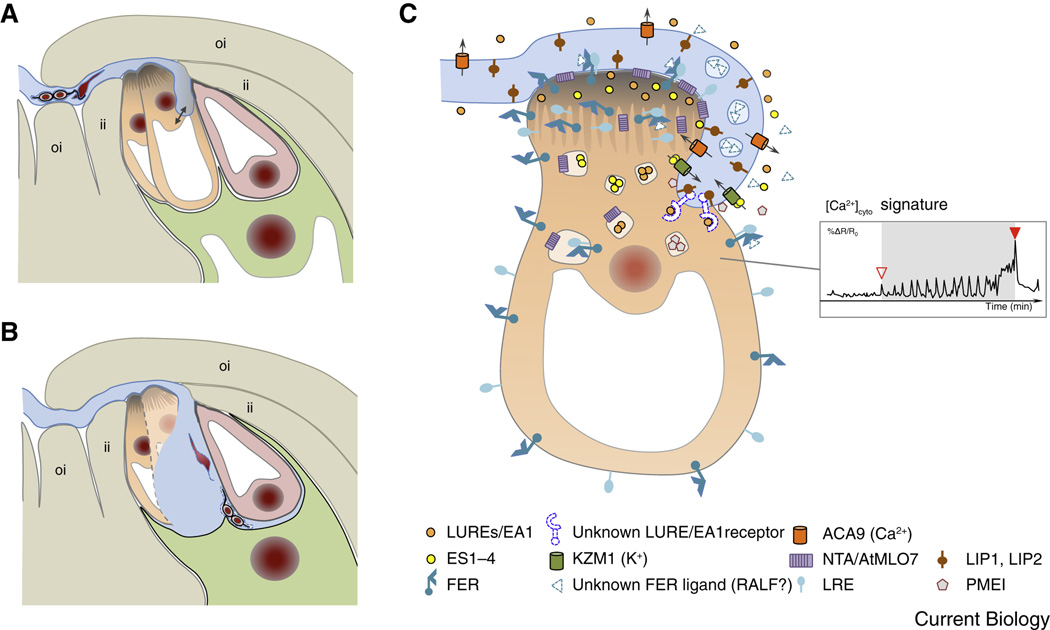
Figure 2. Pollen tube reception involves elaborate communication between the pollen and the receptive synergid cell.
(A) The arriving pollen tube enters the female gametophyte and grows beyond the filiform apparatus, which is formed at the micropylar ends of the synergid cells. The pollen tube continues to grow along or around the receptive synergid cell, extending toward the direction of the central cell. (B) The pollen tube bursts and explosively discharges its contents, including the two immotile sperm cells. Released sperm cells remain physically connected to each other and position between the membranes of the egg cell and the central cell, respectively. (C) Diagram of pollen tube-synergid interactions and the characteristic cytoplasmic (Ca2+)cyto signature induced in the receptive synergid cell. The open triangle in the inset in (C) indicates the moment of first physical interaction between the receptive synergid cell and the pollen tube membrane, the closed triangle marks the moment of pollen tube burst. For details on the molecular events and players of pollen tube-synergid interactions see text. An overview about the molecular players is listed in Suppl. Table S1. Abbreviations: ACA9, Arabidopsis auto-inhibited Ca2+-ATPase 9; EA1, maize EGG APPARATUS1; ES1-4, maize EMBRYO SAC 1-4; FER, Arabidopsis FERONIA; ii, inner integument; KZM1, maize K+ Shaker channel KZM1; LIP, Arabidopsis LOST IN POLLEN TUBE GUIDANCE; LRE, Arabidopsis LORELEI; LUREs, pollen tube attractants of Arabidopsis and Torenia; NTA, Arabidopsis NORTIA; oi, outer integument; PMEI, pectin methyl esterase inhibitor.

