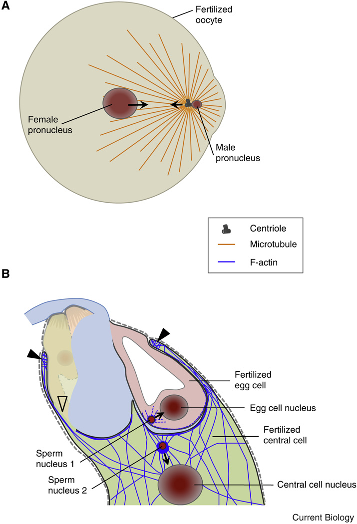
Figure 4. Following plasmogamy the migration of gamete nuclei is realized by microtubules in animals and actin in plants.
(A) Diagram of a fertilized fish oocyte. Sperm aster microtubules are established by the male centrosome. The sperm aster captures the female pronucleus and the two pronuclei rapidly move towards each other and to the center of the oocyte in a dynein-mediated process. (B) In Arabidopsis F-actin dynamics in the female gametes assist the migration of the male nuclei towards the female nuclei while microtubules are not required. After plasmogamy the sperm nucleus in the central cell becomes surrounded by a star-shaped structure of F-actin cables migrating together with the sperm nucleus towards the central cell nucleus. Closed triangles label the characteristic ring-shaped actin network at the micropylar end of the central cell surrounding the synergid cells and the egg cell. Centrioles are not present in plants.

