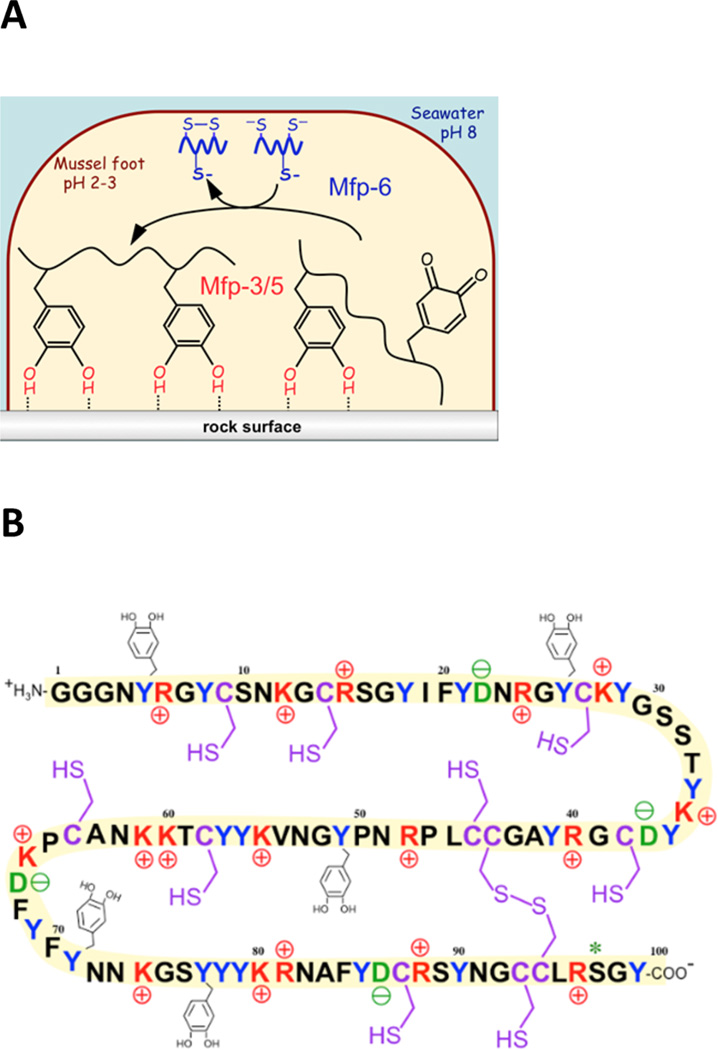
Figure 1.
Mussel foot adhesive proteins. (A) Scheme of the inverted cup configuration of the mussel foot during the initial deposition of adhesive Mfp-3, Mfp-5, and Mfp-6 (which is not adhesive). Oxidized DOPA or Dopaquinone does not contribute to adhesion. (B) Sequence of a mussel foot protein 6 (Mfp-6) variant. Highlighted are basic residues (red), acidic residues (green), tyrosines (blue), and cysteines (purple). Five of 20 tyrosines are shown as being modified to DOPA. Likewise, 2 of 11 cysteines are coupled as disulfide cystine. Although the implied stoichiometry is correct, the specific position of each modification has yet to be determined.

