Fig. 1.
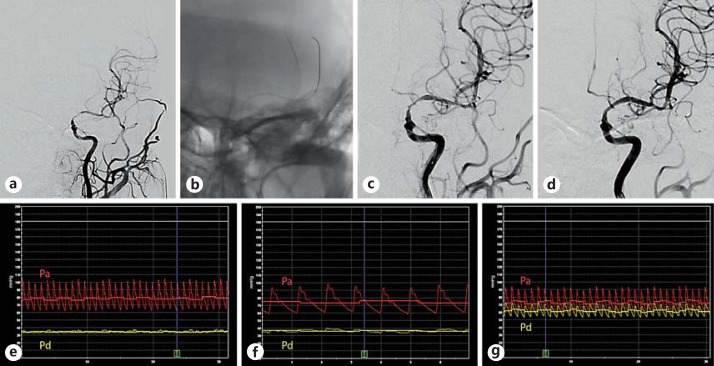
a A patient with a severe stenosis over the left internal carotid artery C7 segment extending to the M1 segment of the middle cerebral artery, with poor opacification of the left anterior cerebral artery on angiography. b Collateral supply via the leptomeningeal branches and the left posterior communicating artery was suboptimal. A pressure wire was advanced 3 cm distal to the stenosis to measure the Pd. As there was significant residual stenosis after initial angioplasty with a 1.5 × 15 mm Gateway balloon (c), repeat angioplasty was performed with a 2.0 × 15 mm Gateway balloon, and a 2.5 × 15 mm Wingspan stent was deployed (d). FF and Pa-Pd gradient were 0.43 and 45 mm Hg before intervention (e), 0.49 and 39 mm Hg after the first angioplasty (f), and 0.84 and 12 mm Hg after final stent deployment (g). The yellow tracing corresponds to Pd, and the red tracing corresponds to Pa. Colors refer to the online version only.

