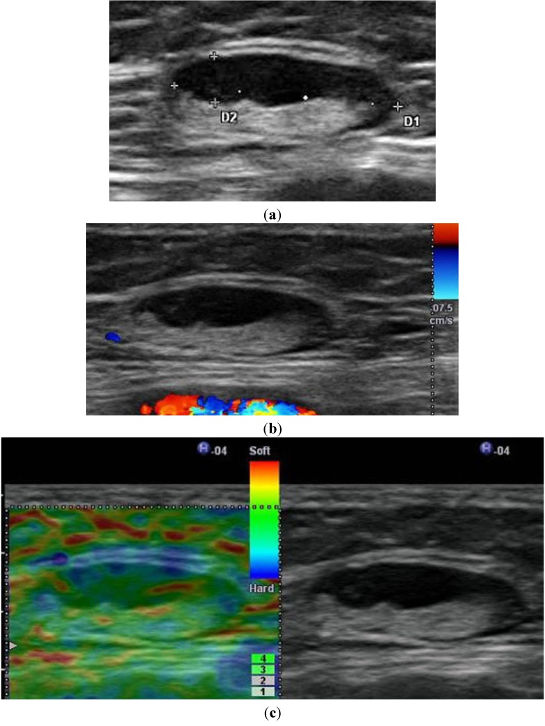
Figure 4.
A 53-year-old woman with cutaneous malignant melanoma of the sole. No lymph node metastasis was confirmed histologically. (a) B-mode US shows a 16.9 × 6.7-mm enlarged lymph node of the inguinal region with an eccentric broadening of the parenchyma and a withdrawal of central echoes to one side (bottom). (b) Color Doppler US shows no central and peripheral perfusion. (c) Elastography shows a mosaic pattern of green and red. The lymph node can be estimated as being soft on the basis of this mosaic pattern. Elastography can provide a benign finding.

