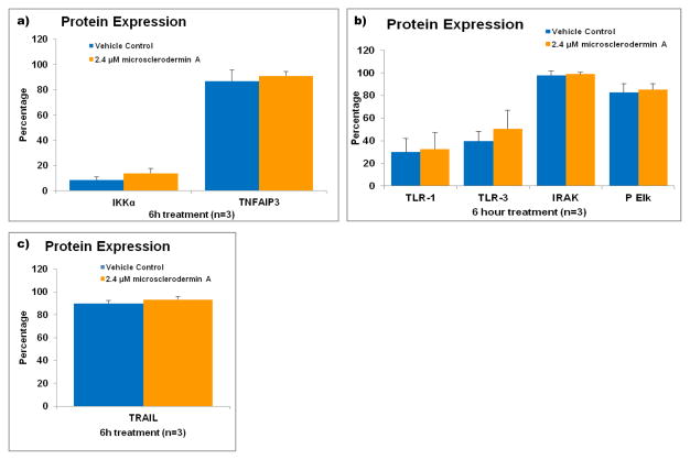
Figure 4. Flow cytometric analysis of pathways involved in the regulation of NFκB in AsPC-1 cells treated with microsclerodermin A.
AsPC-1 cells were treated with either 2.4 μM microsclerodermin A or methanol (vehicle control) for 6 hours and analyzed via flow cytometry to determine changes in expression of a) Iκκα and TNFAIP3, which keep NFκB in its inactive form in the cytoplasm; b) the members of the Toll-like Receptor signaling pathway TLR-1, TLR-3, IRAK and phosphorylated Elk; c) the TNF-related apoptosis-inducing ligand (TRAIL). No significant changes were observed in these pathways in cells treated with microsclerodermin A compared to cells treated with vehicle control. Graphical representation of the flow cytometry data shows the average of 3 experiments. Error bars represent standard deviation.

