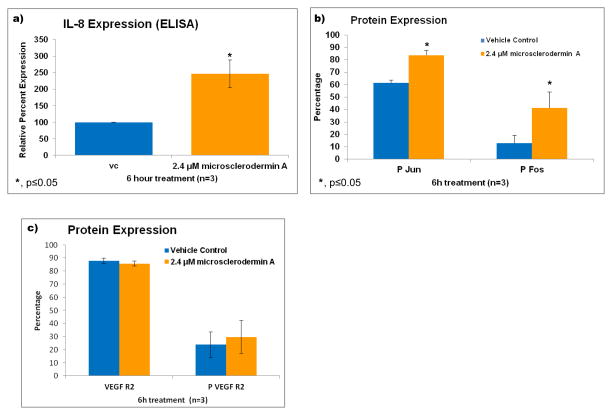
Figure 5. Treatment of AsPC-1 cells with microsclerodermin A increases IL-8 secretion possibly through the upregulation of the transcription factor AP-1, but without affecting VEGF Receptor 2 expression.
a) An increase in IL-8 production by AsPC-1 cells treated with microsclerodermin A for 6 hours compared to control was observed using an ELISA assay. Data shown is the average of 3 experiments ± standard deviation. b) Graphical representation of the flow cytometry data for intracellular staining of AsPC-1 cells treated for 6 hours with 2.4μM microsclerodermin A or vehicle control. Data shown is the average of 3 experiments ± standard deviation. The phosphorylated forms of Fos and Jun were statistically significantly increased in microsclerodermin A treated AsPC-1 cells compared to controls. c) Graphical representation of the flow cytometry data for intracellular staining of AsPC-1 cells treated for 6 hours with 2.4μM microsclerodermin A or vehicle control. Data shown is the average of 3 experiments, error bars represent standard error of the mean. No significant differences in expression of the VEGF receptor 2 in its native and phosphorylated form were observed between control and microsclerodermin A treated AsPC-1 cells.

