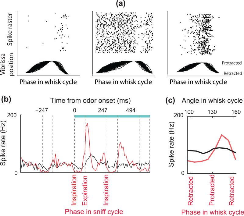
Figure 3. Encoding of odorants and vibrissa contact during the sniffing and whisking cycles.
(a) Whisking solely in air modulates the activity of trigeminal ganglion cells. Unit recordings were obtained from a head-fixed rat together with simultaneous optical tracking of vibrissa position. Vibrissa movements are aligned to protraction onset, retraction onset, and end. Raster plots the spike times above the movements. Adapted from Khatri et al. [19]. (b) Peristimulus time histograms for a mitral/tufted cell in response to an odor stimulus (blue bar, stimulus duration): synchronized by odor onset (black), and temporally warped to the phase in the sniff cycle (red). Note that mitral cell activity is modulated by sniffing before the odor presentation, and that odor-induced responses are more tightly time-locked to the sniff phase than to the time after odor onset. Vertical dashed lines indicate the beginning and end of inhalation intervals. Adapted from Shusterman et al. [21]. (c) Touch response in vibrissa sensory cortex is strongly modulated by the phase in the whisk cycle. The red trace shows the spike rate of a neuron in response to object contact parsed according to the phase in the whisk cycle at which the contact occurred. The black line is the same data parsed according to the angular position of the vibrissa where the contact occurred. Unlike the case for phase, there is no significant tuning for angle. Adapted from Curtis and Kleinfeld [24].

