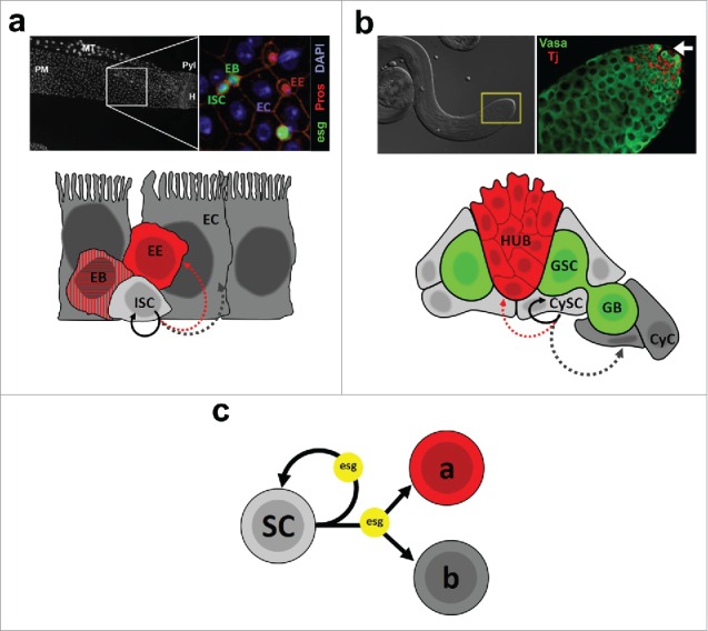
Figure 1.

(a) The Drosophila midgut epithelium. Top left: DAPI nuclear staining of the posterior midgut (PM). MT: Malpighian tubule; Pyl: Pyloric ring; H: Hindgut. Top right: Immunostaining of a region that corresponds approximately to the area depicted in the left image. ISC/EBs are labeled by expression of an esg-GFP reporter (esg), EEs are identified by Prospero (Pros) nuclear staining, and ECs are identified based on their polyploid nuclei (DAPI). Images by Christopher Koehler, L. Jones lab. Bottom: Cartoon representing the 4 cell types that make up the midgut epithelium and their lineage relationships. ISCs can self-renew (solid arrow) or differentiate into EEs or ECs (dashed arrows - see main text for details). (b) The Drosophila testis. Top left: DIC image of a testis. The inset marks the apical tip of the gonad, where the stem cell niche resides. Top right: Immunostaining of a testis tip. Germ cells are identified based on expression of Vasa, whereas somatic cells are labeled by expression of Traffic jam (Tj). The arrow points to the approximate location of the apical hub (not shown). Bottom: Schematic representation of the testis apical tip, showing somatic hub cells, germline and somatic stem cells (GSCs and CySCs, respectively) and their progeny (GB and CyC, respectively). Lineage relationships are shown only for CySCs, which can self-renew or differentiate into CyCs or, more rarely, give rise to hub cells. (c) Abstract synthesis of our preliminary work on the role of escargot (esg), which is simultaneously required for the maintenance of stem cells (SC - testis CySCs 2 and midgut ISC/EBs 4), while also controlling the differentiation of their progeny into alternative fates (“a” and “b”). 1,4
