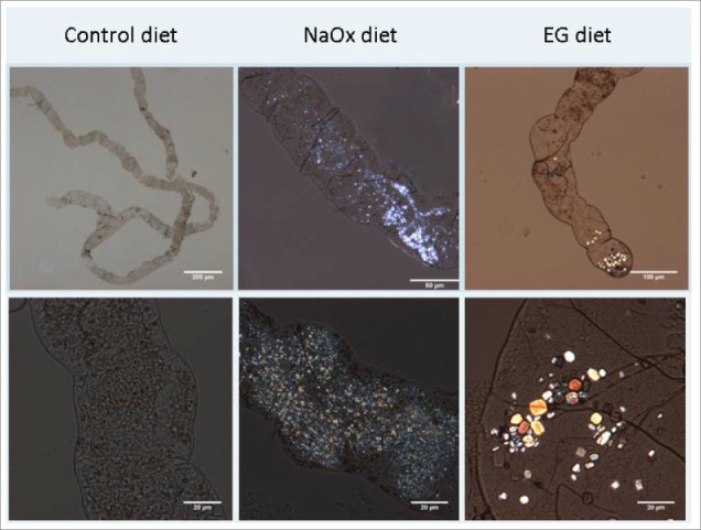
Figure 1.

Crystal formation in Malpighian tubules 10-40x magnification. The control diet showed the normal structure of a pair of Malpighian tubules joining into a single ureter, which further secretes into hindgut. The crystal morphology was small and extensive in the NaOx (sodium oxalate) treatment group. In contrast EG (ethylene glycol) enriched diet induced crystals which were bigger, poly-angular with a jewel-like gloss.
