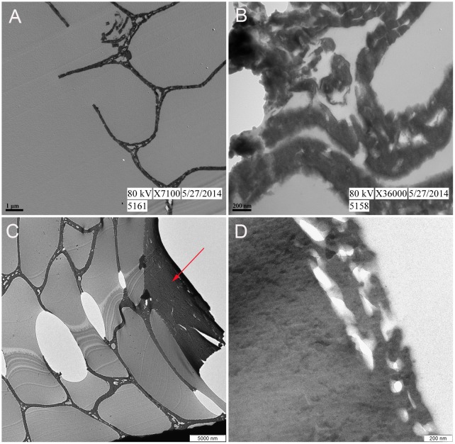
Fig 4. TEM images of 350°C feather fragments.
(A-B) Unidentifiable piece of 350°C feather at lower and higher magnification, respectively. The honey-comb structure observed in (A) indicates it is the pith of either a barb or rachis. (C-D) Feather fragment positively identified as barb. (C) External cortex (arrow) and internal pith are observed. (D) At higher magnification no electron dense microbodies consistent with melanosomes are observed in the cortex.

