
FIG 5.
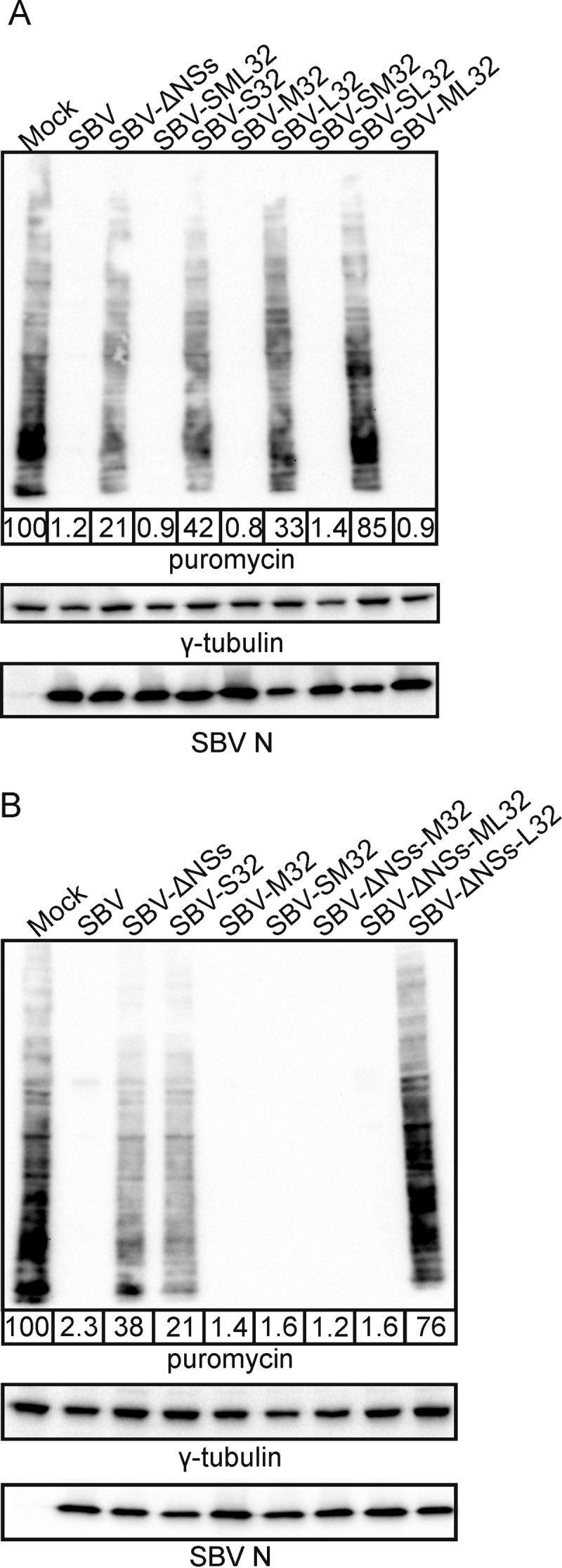
The M segment of SBVp32 rescues the inability of attenuated S segments to induce total cellular protein shutoff. Western blots display puromycin-labeled proteins 16 h after infection with the indicated reassortants (MOI of 1). γ-Tubulin was used as a loading control, and SBV N was used to confirm infection. The numbers indicate the quantification of protein levels in each lane relative to that for mock infection, which was set at 100%. (A) Reassortants carrying the S segment of SBVp32 in combination with wild-type or SBVp32 M and L segments. (B) Reassortants carrying a wild-type SBV S segment deleted of its NSs protein in combination with wild-type or SBVp32 M and L segments.