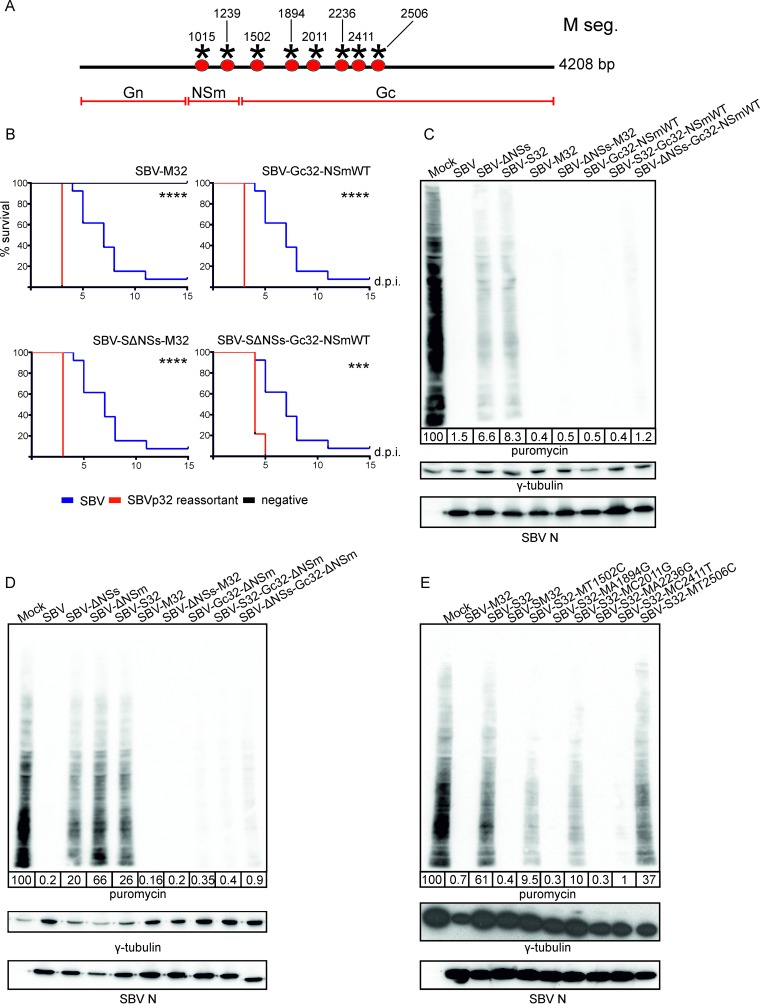
FIG 6.
SBVp32 pathogenicity maps to the Gc glycoprotein. (A) Schematic representation of SBVp32 mutations within the M segment. (B) Survival plots of 8-day-old NIH-Swiss mice inoculated intracerebrally with 400 PFU of the indicated reassortants. Asterisks indicate significance levels (***, P ≤ 0.001; ****, P ≤ 0.0001 [as determined by a log rank test and a Mantel-Cox test). (C to E) Western blots displaying puromycin-labeled proteins 16 h after infection with the indicated reassortants (MOI of 1). γ-Tubulin was used as a loading control, and SBV N was used to confirm infection. The numbers indicate the quantification of protein levels in each lane relative to that for mock infection, which was set at 100%.

