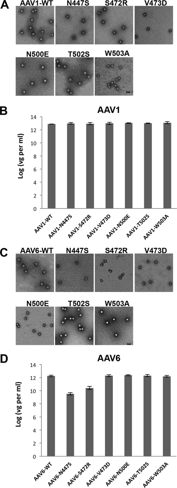
FIG 2.

WT AAV and SIA-binding site mutant characterization. (A and C) Negative stain micrographs and (B and D) genome packaging titers of WT AAV and SIA binding site mutants. Panels A and B show results for AAV1, and panels C and D show results for AAV6. The genome packaging titers were determined by q-PCR. The values are means from three experiments displayed in a log scale; the error bars represent standard deviations. vg, vector genome.
