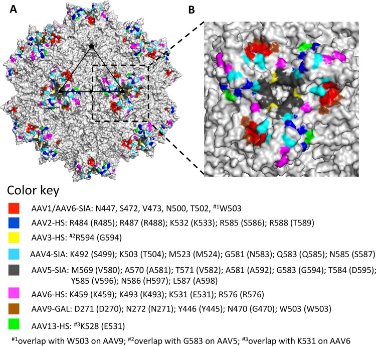
FIG 8.
AAV glycan receptor binding footprints. (A) Available glycan binding sites for AAV1, AAV2, AAV3B, AAV4, AAV5, AAV6, AAV9, and AAV13 projected onto the surface density of the AAV1 capsid. (B) Closeup of the receptor binding footprints viewed looking down the icosahedral 3-fold axis. Residues involved in glycan interactions for the different AAV serotypes are listed in the color key. Residues in parentheses indicate AAV1 numbering. The figures were generated using the PyMOL program (52).

