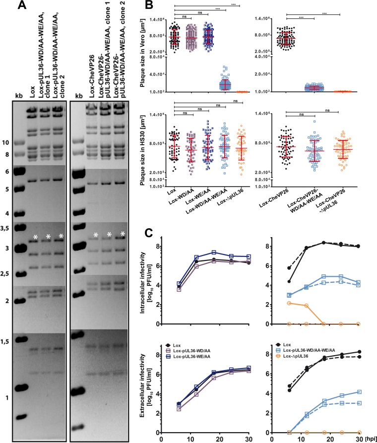
FIG 2.
Characterization of HSV-1-pUL36 mutants. (A) Agarose gels of AscI restriction digests of the parental BACs pHSV-1(17+)Lox and pHSV-1(17+)Lox-mCheVP26 or the derived mutants pHSV-1-pUL36-WD/AA-WE/AA and pHSV-1-mCheVP26-pUL36-WD/AA-WE/AA. The sizes of the restriction fragments and molecular size markers are indicated in kilobase pairs. (B) Plaque formation in Vero and Vero-HS30 cells. The plaque sizes in Vero cells were determined in three independent experiments and those in Vero-HS30 cells in two independent experiments. Values are means ± standard deviations (SD) (***, P < 0.0001 as determined by Tukey's multiple comparison test). (C) Vero cells were infected with an MOI of 5 PFU/cell (1 × 10⁷ PFU/ml), and the samples were harvested at the indicated time points. The extracellular and intracellular infectivities were determined in duplicates. The virus yields of the parental strains Lox (solid line) and Lox-CheVP26 (dashed line) are depicted by closed circles, those of Lox-WD/AA (solid dark blue line), Lox-WE/AA (solid blue line), Lox-WD/AA-WE/AA (solid light blue line), and Lox-CheVP26-WD/AA-WE/AA (dashed light blue line) by squares, and those of the deletion mutants HSV-1-ΔUL36 deletion mutants are represented by open circles with a solid or dashed line for Lox and Lox-CheVP26, respectively.

