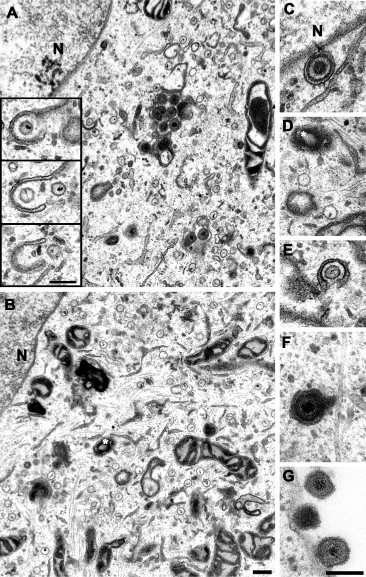
FIG 4.
Accumulation of cytosolic capsids and reduced secondary envelopment in cells infected with HSV-1(17+)Lox-pUL36-WD/AA-WE/AA. Vero cells were infected synchronously with HSV-1(17+)Lox (A) or HSV-1(17+)Lox-pUL36-WD/AA-WE/AA (B to G) PFU, fixed at 14 hpi, and analyzed by electron microscopy. The inset in panel A contains three consecutive sections showing viral genomes within cytoplasmic capsids. A white asterisk indicates the microtubule-organizing center, and N indicates the nucleus. The primary enveloped virion (C), cytosolic capsids (D), wrapping intermediate (E), enveloped virus particle (F), and extracellular virions (G) were assembled upon infection with HSV-1-pUL36-WD/AA-WE/AA. Scale bars, 200 nm.

