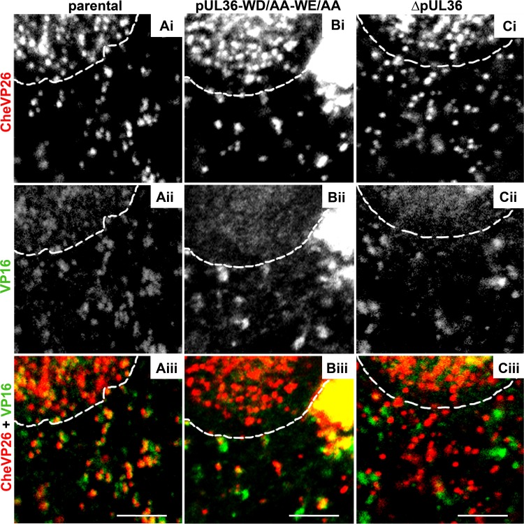
FIG 7.
The capsids of HSV-1-CheVP26-pUL36-WD/AA-WE/AA acquire VP16. Vero cells were infected synchronously with HSV-1(17+)Lox-CheVP26 (A), HSV-1(17+)Lox-CheVP26-WD/AA-WE/AA (B), or HSV-1(17+)Lox-CheVP26-ΔUL36 (C) and fixed at 10 hpi. The specimens were labeled for VP16 (pAb 631209; white in panels ii and green in panels iii) and analyzed by confocal microscopy. The capsids were visualized by CheVP26 (white in panels i and red in panels iii). The nuclei were identified by DAPI labeling and are depicted by dashed white lines. Scale bars, 3 μm.

