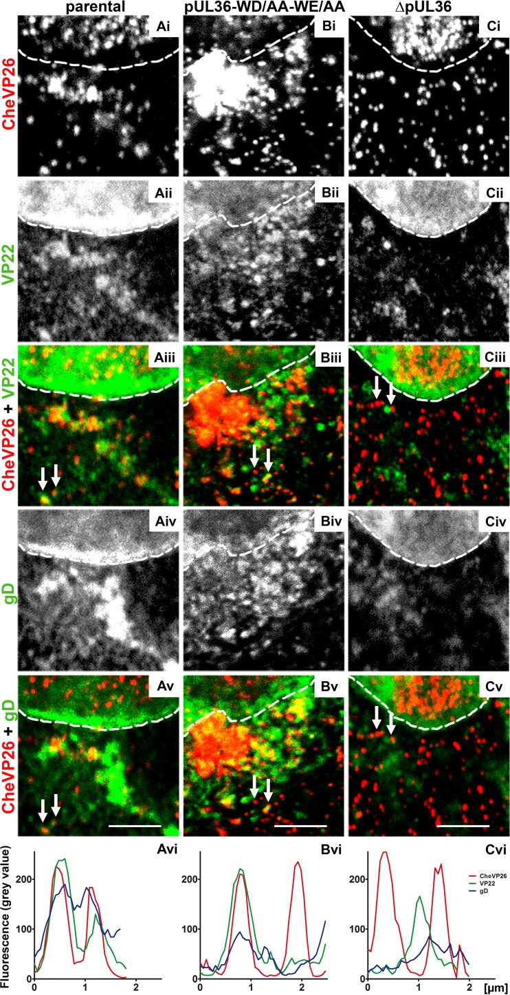
FIG 8.
Few capsids of HSV-1(17+)Lox-mCheVP26-pUL36-WD/AA-WE/AA undergo secondary envelopment. Vero cells were infected synchronously with HSV-1(17+)Lox-mCheVP26 (A), HSV-1(17+)Lox-mCheVP26-pUL36-WD/AA-WE/AA (B), or HSV-1(17+)Lox-CheVP26-ΔUL36 (C), fixed at 10 hpi and labeled for VP22 (pAb AGV30; white in panels ii and green in panels iii) and glycoprotein D (MAb DL-6; white in panels iv and green in panels v) and analyzed by confocal fluorescence microscopy. The capsids were detected by mCheVP26 (white in panels i and red in panels iv and v). The nuclei were identified by DAPI labeling and are depicted by dashed white lines. The fluorescence intensity of randomly selected capsids (white arrows) was measured in 8-bit images along a 1-pixel-thick and 2- to 2.5-μm-long line. The histograms represent the plotted gray values of CheVP26 (red), VP22 (green), and gD (blue) against the length of the line in micrometers. Scale bars, 3 μm.

