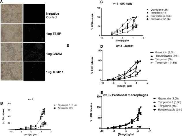
Fig 4. Temporizin-1 possesses low toxicity against mammalian cells of diverse species.

A: J774 cells were incubated at 37°C for 60 minutes for a cell permeabilization assay. A single dose of 1 μg/ml was applied to compare the efficiency of the gramicidin (A2), temporizin (A3), gramicidin (A4) and temporizin-1 (A5) peptides to injure the J774 cell line. B: Dose-response relationship of temporizin and temporizin-1 after treatment for 60 and 90 minutes, respectively on mouse J774.G8 cell lineage. The cytotoxic actions was quantified by LDH release assay. C: A graphic representing LDH release performed in human Jurkat cells after adding the peptides and benznidazole at different concentrations. D: A graphic representing LDH release performed in the rat adenoma GH3 cell line after adding the peptides and benznidazole at different concentrations. E: A graphic represention of the LDH Release Assay that was performed after adding the peptides and benznidazole at different concentrations to primary mice peritoneal macrophages The values represent the mean ± SD of three to five experiments (cell permeabilization assay) performed on different days.
