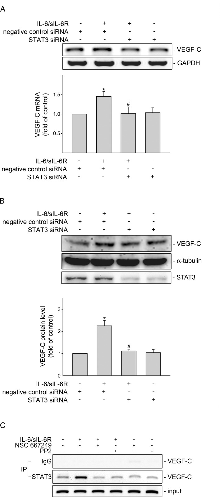
Fig 4. STAT3 siRNA suppressed IL-6/sIL-6R-induced VEGF-C expression in SV-LECs.
(A) Cells were transiently transfected with negative control siRNA or STAT3 siRNA for 48 h. After transfection, cells were treated with IL-6 plus sIL6R (20 ng/ml) for another 6 h. The extent of VEGF-C mRNA was analyzed by RT-PCR as described in the “Materials and Methods” section. Each column represents the mean ± S.E.M. of five independent experiments. *p<0.05, compared to the control group; # p<0.05, compared to the vehicle-treated group in the presence of IL-6 plus sIL6R. (B) Cells were transiently transfected with negative control siRNA or STAT3 siRNA for 48 h. After transfection, cells were treated with IL-6 plus sIL6R (20 ng/ml) for another 24 h. The VEGF-C protein level was then determined by immunoblotting. Each column represents the mean ± S.E.M. of four independent experiments. * p<0.05, compared to the control group; # p<0.05, compared to the vehicle-treated group in the presence of IL-6 plus sIL6R. (C) Cells were pretreated with the vehicle, PP2 (1 μM) or NSC 667249 (0.3 μM) for 30 min followed by treatment with IL-6 plus sIL6R (20 ng/ml) for another 4 h. The ChIP assay was performed as described in the ‘‘Materials and methods” section. Typical traces representative of three independent experiments with similar results are shown. The VEGF-C promoter region (-367/-198) was detected in the cross-linked chromatin sample before immunoprecipitation (bottom panels of the chart, Input, positive control).

