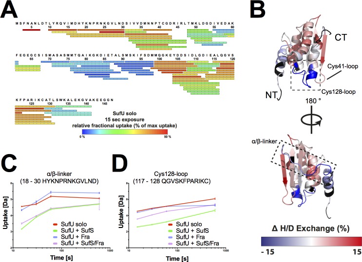
Fig 4. BsSufS alters the H/D exchange of BsSufU upon binding.
(A) Detected peptic peptides of BsSufU with the relative fractional uptake after 15 s of incubation in deuterated buffer. (B) Changes in the relative fractional deuterium uptake of BsSufU after incubation with BsSufS for 15 s in D2O buffer compared to BsSufU alone were mapped onto the surface of BsSufU (PDB ID 2AZH). The heat map represents the differences in deuterium uptake compared to BsSufU alone. A decrease (blue) in deuterium uptake signals protection (i.e., a binding event), whereas an increase (red) signals a structural rearrangement. Black regions were not detected. Binding of BsSufU to (C) the α/β-linker and (D) the Cys128-loop of BsSufS as a function of deuterium uptake over time. Color code: BsSufU alone (red), BsSufU + BsSufS (green), BsSufU + BsFra (blue), and BsSufU + BsSufS/BsFra (violet). N-terminus (NT) and C-terminus (CT).

