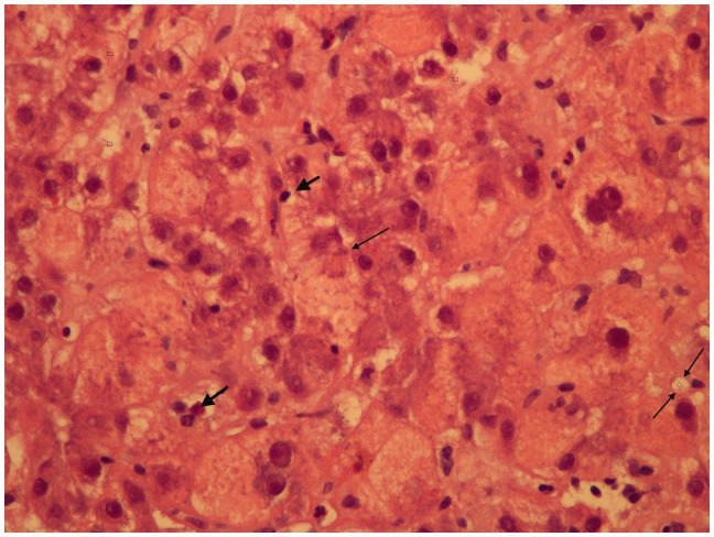
Figure 1.
The hepatocytes show vacuolization of their cytoplasm and focal cytoplasmic expansion, i.e. ballooning degeneration, (single thin arrow) indicating trans-membrane transport mechanisms are breaking down and scattered necrotic hepatocytes. This appearance is quite distinctive from fatty liver of pregnancy, where the hepatocytes would show a uniform microvescicular and foamy appearance. Intracanalicular cholestasis is present (two thin arrows). There are some neutrophilic leukocytes responding to cell death. There is a light patchy sinusoidal lymphocyte infiltrate (thick arrows)—but the appearance is not typically auto-immune, where a more intense lymphoplasmacytic infiltrate would be expected. This appears to be more of a direct toxicity to the hepatocytes.

