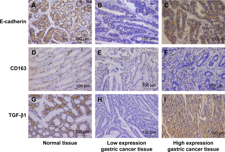
Figure 1.
Immunohistochemical results of E-cadherin, TGF-β1, and CD163 in the gastric cancer and paracancer tissue.
Notes: (A) Typical high expression of E-cadherin in paracancer tissue of gastric cancer. Staining was localized predominantly in the cytomembrane. (B) Typical low expression of E-cadherin in gastric cancer tissue. (C) Infrequent high expression of E-cadherin in gastric cancer tissue. (D) Low expression of CD163 in normal tissue. Staining was localized predominantly in the cytosol. (E) Low expression of CD163 in gastric cancer tissue. (F) High expression of CD163 in gastric cancer tissue. (G) Low expression of TGF-β1 in paracancer tissue. Staining was localized predominantly in the cytosol. (H) Low expression of TGF-β1 in gastric cancer. (I) High expression of TGF-β1 in gastric cancer. Magnification, 200×.
Abbreviation: TGF-β1, transforming growth factor-β1.

