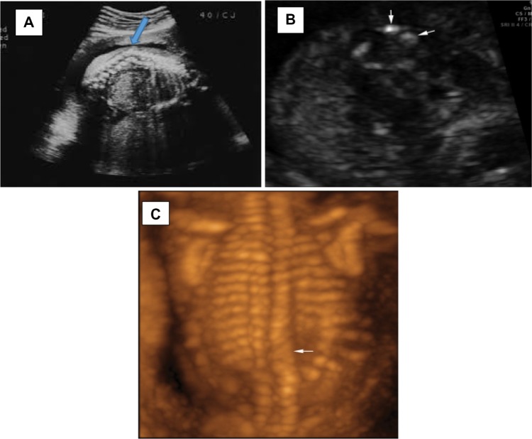
Figure 1.
Prenatal diagnosis of Alagille syndrome.
Notes: (A) A prenatal anatomy scan revealing kyphosis with underlying thoracic vertebral anomaly (blue arrow). (B) A transverse image shows thoracic hemivertebrae (arrows). (C) A three-dimensional sonogram in the posterior–anterior plane demonstrates classic butterfly and hemivertebrae (arrow).

