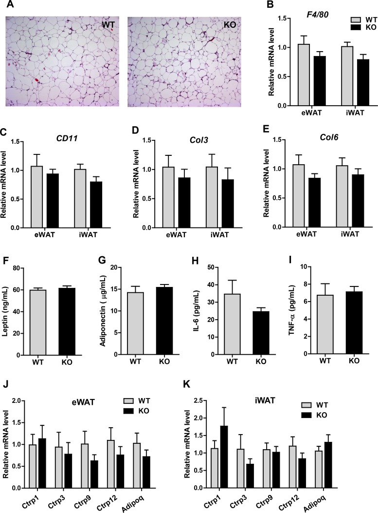
Fig. 9.
Inflammatory and fibrotic states of adipose tissue in Ctrp5-null mice. A: representative histological sections of eWAT from WT and KO mice stained with hematoxylin and eosin. B and C: quantitative PCR analysis of macrophage marker genes (F4/80 and Cd11) in visceral (epididymal; eWAT) and subcutaneous (inguinal; iWAT) white adipose tissue. D and E: expression levels of fibrotic collagen genes (Col3 and Col6) in the visceral (eWAT) and subcutaneous (iWAT) fat depots of WT and KO mice. F–I: ELISA quantification of serum leptin, adiponectin, IL-6, and TNFα levels in WT and KO mice. J and K: expression levels of adiponectin and CTRPs in the eWAT and iWAT of WT and KO mice. All expression levels were normalized to 18s rRNA levels. WT, n = 8; KO, n = 7.

