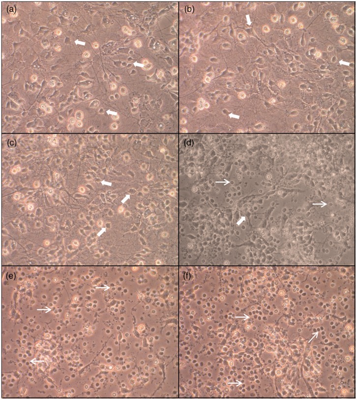
Figure 1.
Morphological changes and death in rat brain cells exposed to A549 conditioned media. Phase-contrast microscopic observation (×20) of primary rat mixed cortical cultures 24 h after treatment with 2% (b), 5% (c), 15% (d), 25% (e), or 50% (f) A539 conditioned medium as well as controls (a). Thick arrows show normal viable neuronal bodies while thin arrows show round damaged or dead cells. See text for details. (A color version of this figure is available in the online journal.)

