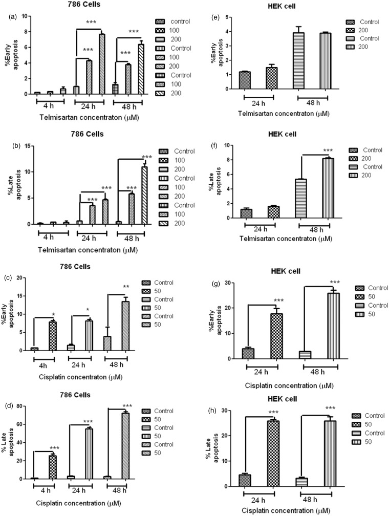
Figure 3.
Apoptosis induced by telmisartan and cisplatin in 786 RCC cells was detected using flow cytometry. Levels of early apoptosis (EA) (a) and late apoptosis (LA) (b) were detected for cells treated with 100 µM and 200 µM telmisartan for 4 h, 24 h, and 48 h. Levels of EA (c) and LA (d) were also detected for cells treated with 50 µM cisplatin for 4 h, 24 h, and 48 h. Apoptosis induced by telmisartan and cisplatin in HEK cells was detected using flow cytometry. Levels of EA (e) and LA (f) were detected for cells treated with 200 µM telmisartan for 24 h and 48 h. Levels of EA (g) and LA (h) were detected for cells treated with 50 µM cisplatin for 24 h and 48 h. ***P < 0.001; **P < 0.01; *P < 0.05

