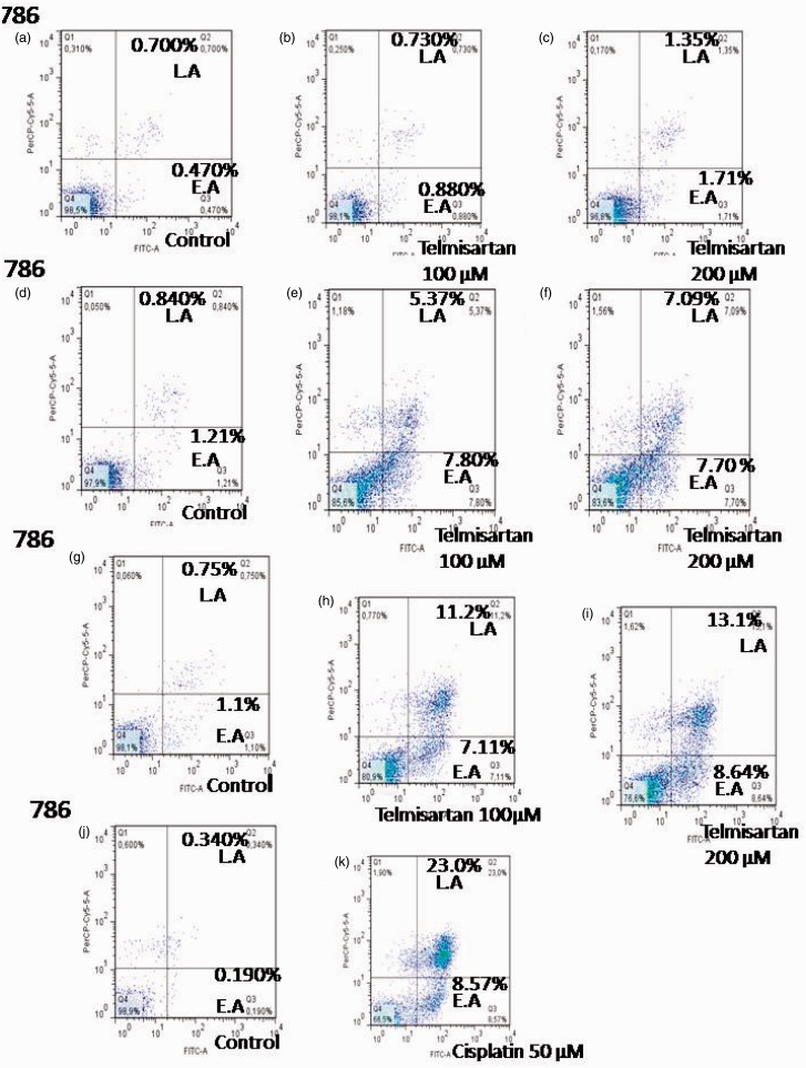
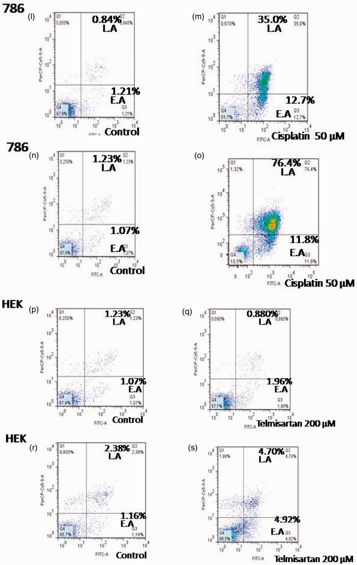
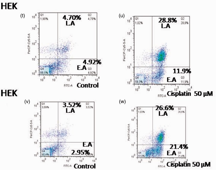
Figure 4.
Effects of telmisartan and cisplatin on early and late apoptosis as detected using flow cytometry. Treatment of 786 RCC cells with 100 µM and 200 µM telmisartan induced early- or late-stage apoptosis after 24 h (e, f) and 48 h (h, i), but not at the 4 h timepoint (b, c). In contrast, treatment of normal HEK cells with 200 µM telmisartan did not induce apoptosis after 24 h (q), yet did after 48 h (s). The lower right quadrants represent the Annexin V-FITC-positive/PI-negative cells in the early stages of apoptosis, while the top right quadrants include Annexin V-FITC-positive/PI-positive cells in the late stages of apoptosis. Treatment with 50 µM cisplatin induced either early or late apoptosis in 786 RCC cells at the 4 h (k), 24 h (m), and 48 h (o) timepoints, and a greater number of late stage cells were observed. Treatment with 50 µM cisplatin also induced early and late apoptosis in HEK cells 24 h (u) and 48 h (x) after treatment, with the latter being more predominant. (A color version of this figure is available in the online journal.)


