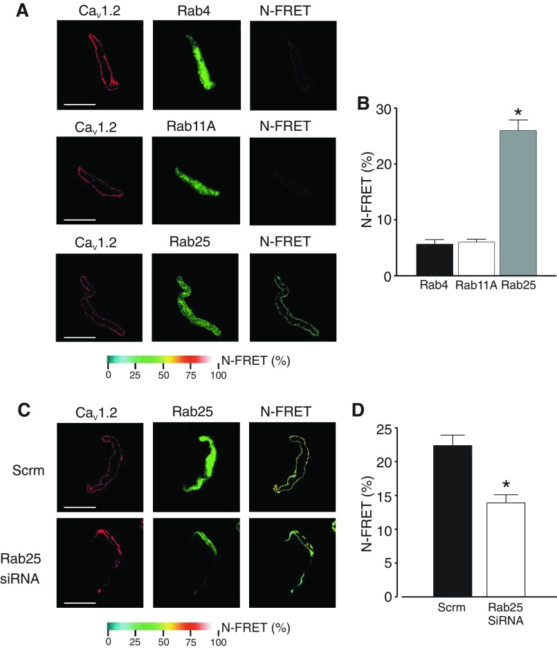
Fig. 2.
Rab25 colocalizes with CaV1.2 channels and stimulates CaV1.2 surface expression. A: representative images of myocytes labeled with fluorescent antibodies to either Rab4, Rab11A, or Rab25 (Alexa546) or CaV1.2 (Alex488) and respective normalized fluorescence resonance energy transfer (N-FRET) images. B: mean N-FRET data (n = 10). C: representative images of myocytes isolated from arteries treated with either scrm or Rab25siRNA labeled with fluorescent antibodies to either Rab25 (Alexa546) or CaV1.2 (Alex488) and respective N-FRET images. D: mean N-FRET data (n = 10). *P < 0.05, compared with scrm control. Scale bars = 10 μm.

