Fig. 3.
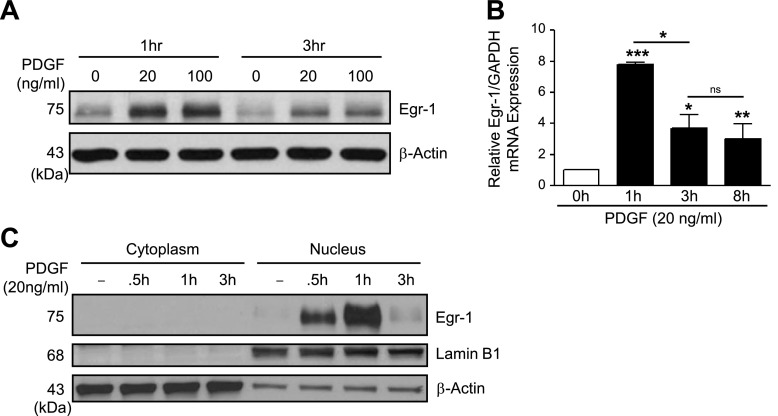
PDGF increases EGR1 expression in hPASMCs. A: representative Western blotting images demonstrate increased EGR1 protein expression in human pulmonary artery smooth muscle cells (hPASMC) following stimulation with PDGF-BB (20–100 ng/ml, 1–3 h). B: EGR1 mRNA expression levels are increased in hPASMC following PDGF-BB treatment (20 ng/ml, 0–8 h) relative to GAPDH expression. C: Western blotting with cytoplasmic and nuclear hPASMC fractions demonstrates PDGF-BB treatment (20 ng/ml, 0–3 h) induces EGR1 expression limited to the nucleus, with Lamin B1 used as a nuclear loading control. Results are shown as means ± SE from at least 3 experiments. *P < 0.05, **P < 0.01, ***P < 0.001 vs. control unless otherwise indicated.

