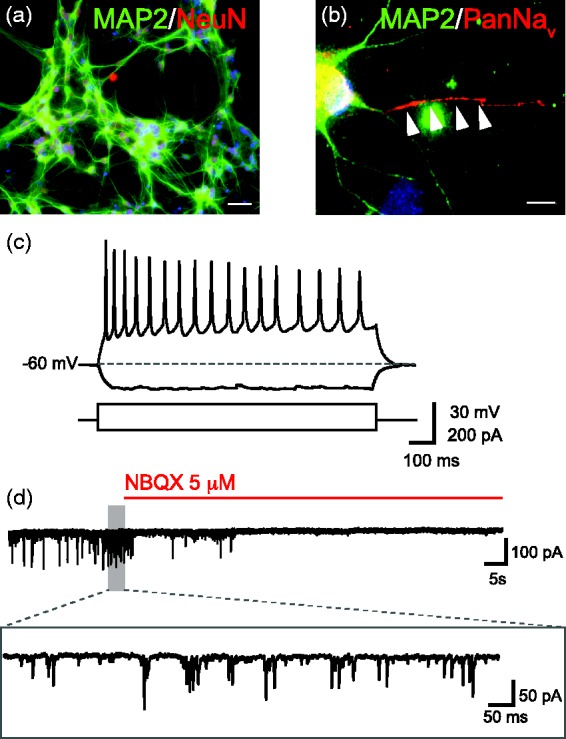
Figure 1.

Morphology and electrophysiological properties of cultured Ngn2 induced neurons. (a) Representative image of MAP2/NeuN double immunostaining of Ngn2-infected iPSC-derived iNs. (b) High-magnification image of a pan-NaV antibody staining correctly localized at the axonal shaft of a Ngn2-iN. In (a) and (b), Hoechst is used as nuclear counterstaining (blue). Scale bar, 50 μm in (a) and 10 μm in (b). (c) Voltage change in response to injection of positive and negative current pulses (+60 and −40 pA, respectively). (d) Spontaneous synaptic activity before and after extracellular perfusion with the AMPA-receptor antagonist NBQX (5 μM). The portion of the trace included in the grey area is magnified below. (A color version of this figure is available in the online journal.)
