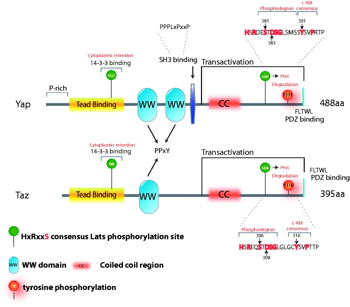
Figure 1.
Schematic representation of the WW proteins Yap and Taz. Yap and Taz structural features, including the WW domains, Tead binding domain, coiled-coil region, and proline-rich and PDZ-binding domains are indicated. Phosphorylation sites described in the text are depicted. The Yap Y391 site is the same as Y357 in the Yap isoform with one WW domain. The sequences encompassing the phosphodegrons are shown. (A color version of this figure is available in the online journal.)

