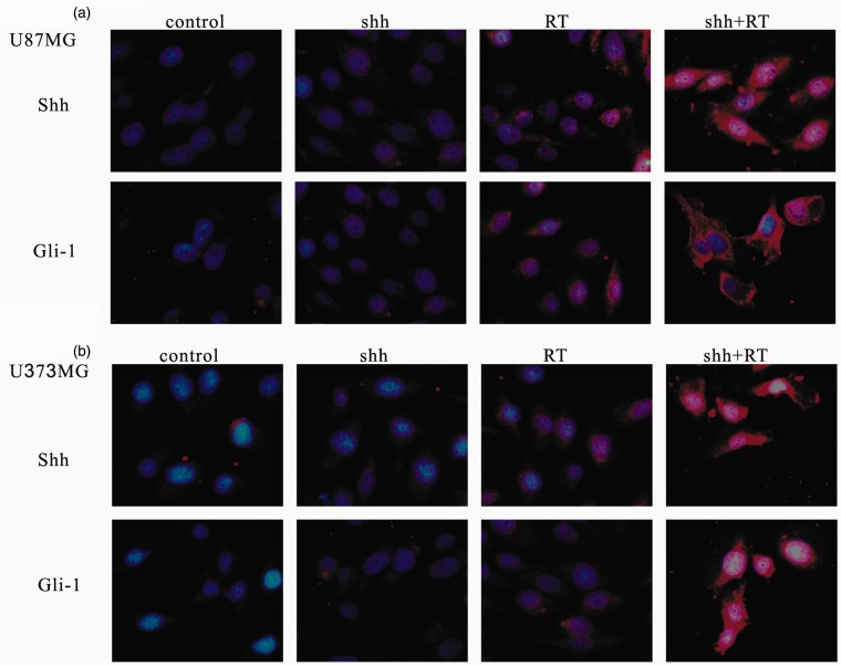
Figure 1.
Immunofluorescence analysis for expression of Shh and Gli-1 in human glioblastoma cells. (a) U87MG cells. (b) U373MG cells. For immunofluorescence staining, cells were harvested after radiation, stained with Shh or Gli-1 antibodies and secondary Rhodamine Red-conjugated antibody (red), and nuclei were counterstained with DAPI (blue). Original magnification 400×

