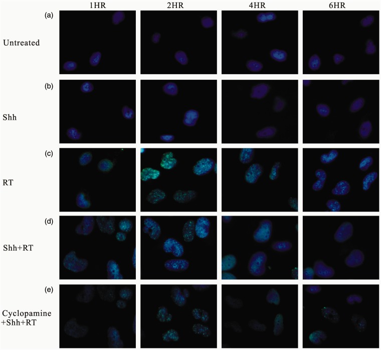
Figure 8.
Immunofluorescent staining for expression of gamma-H2AX in glioblastoma cells with WOX1 overexpression. Cells that were (a) untreated (control) or treated with (b) Shh ligand, (c) radiation of 2 Gy, (d) Shh ligand plus radiation, and (e) cyclopamine plus Shh ligand and radiation. For immunofluorescent staining, cells were harvested after radiation, stained with gamma-H2AX antibody, secondary Cyt-2-conjugated donkey anti-mouse IgG (green), and nuclei were counterstained with Hoechst 33342 (blue). Original magnification 400×

